How Cardio-HART Works?
Cardio-HART is a 6-minute cardiac test that works just like an ECG—only smarter.
How Cardio-HART Works in Practice
Same workflow as today — just one extra sensor
| Step | What staff do now with an ECG | What changes with CHART | Time impact |
|---|---|---|---|
| Apply electrodes | 10 standard leads on chest & limbs | Exactly the same 10 leads | 1 min 20 sec |
| Attach device | Clip ECG cables Place Sensor | Clip ECG cables place a thumb-size chest patch (PCG + MCG microphone/accelerometer) | 20 sec |
| Press Start & wait | Recording | Three 1-minute recordings captured automatically | 3 min 20 sec |
| Print/Save report | AI interpretation cloud processing | Dual report: (1) full ECG analysis, (2) echo-equivalent findings—including LVEF, valve grades, LV mass | 1 min |
No sonographer needed, no cardiologist for reporting, no new room set-up.
If your nurse or HCA can run an ECG, they can run CHART—total bed time ≈ 6 minutes.
What’s inside the box?
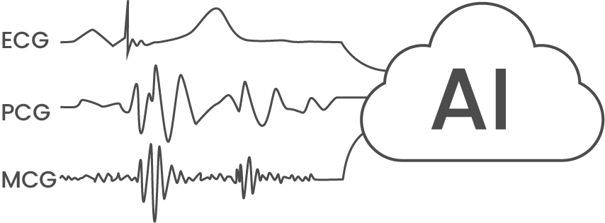
Synchronous tri-modal capture
- ECG: electrical activation
- PCG: valve acoustics (heart sounds)
- MCG: chest-wall micro-motion (surrogate stroke volume)
All three streams share a common clock, letting the AI track electro-mechano-acoustic coupling beat-by-beat.


Multimodal AI fusion
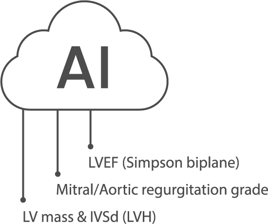
Trained on > tens of thousands of echo-paired patients, a convolutional-transformer ensemble converts the raw signals into:
| Echo-equivalent output | Validation vs. reference echo* |
|---|---|
| LVEF (Simpson biplane) | ± 5 % (95 % LoA), r = 0.89 |
| Mitral/Aortic regurgitation grade | κ = 0.80 |
| LV mass & IVSd (LVH) | Sens 92 % / Spec 90 % |
*Multi-centre cohort; full tables in downloadable white paper.
Key enablers
- Beat-to-beat averaging over 60 s removes most biological and motion noise that plague 10 s ECGs.
- Echo-ground-truth training (no ECG label bias).
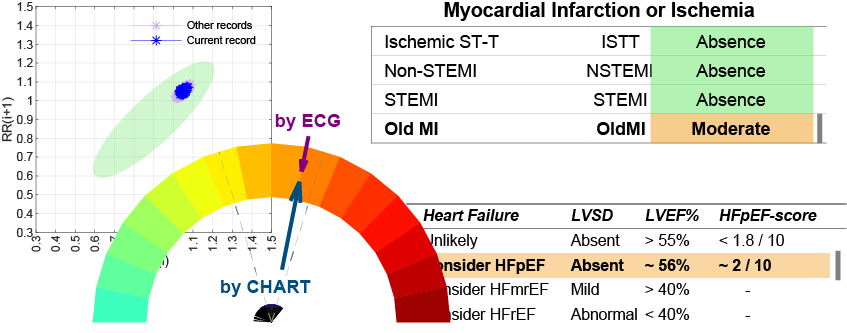
- Explainable reports: numeric values + traffic-light triage + signal quality score.

What it means in practice
Cardiologists
- Echo-level insight before the patient arrives—prioritise scanning slots for true positives.
- Full signal export for audit; AI explanations mapped to guideline criteria.
GPs & Primary Care
- One test answers: Treat now? Refer routinely? Fast-track?
- 94 % drop in “uncertain ECG” flags in pilots.
- No sonographer backlog; CHART printout uses the same EMIS/SystmOne import path as ECG.
Why It Matters Beyond the Clinic

Understanding how Cardio-HART works is more than a technical detail—it shows why it changes outcomes across the healthcare system. By combining ECG, PCG, and MCG into one synchronised test, the system reduces uncertainty, speeds up decision-making, and ensures patients are managed correctly from the very first appointment. This efficiency not only benefits clinicians but also reduces hospital bottlenecks, avoids unnecessary referrals, and gives patients peace of mind. In real-world pilots, Cardio-HART has already demonstrated faster heart failure detection and improved referral accuracy, proving that smart integration of AI can deliver both clinical and economic impact.
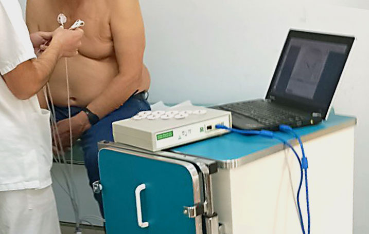
Sequence of event. Quick, non-invasive, immediate reporting
Patient is readied.
ECG electrodes placed and connected.
PMCG sensor placed on thoracic wall between electrode 3 and 4 and holding strap secured.
Recording, 3 x 1 minutes (moments of Zen). Finished, tests are uploaded to cloud for AI processing while patient gets dressed.
Reports typically available before the patient leaves the examination room. (dependent on internet speed).

Next steps?
Same test—one extra sensor, two extra minutes, echo-quality answers.
Cardio-HART is a direct substitute for ECG in clinical practice—only smarter!
